Peter So, PhD

Research Highlights
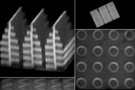
Our laboratory creates novel optical imaging, manipulation, and fabrication technologies on the microscopic level to advance biological sciences and to improve medical practices.
Research:
Many advances in biology and medicine are driven by the availability of new diagnostic tools. Our research focuses on the engineering of novel microscopy instrumentation and the application of these new tools to study biomedical problems. The problems tackled in my laboratory range from understanding the structure/function of single proteins, nature’s smallest machines, to the development of a new non-invasive optical method for cancer diagnosis.
Biography:
Professor So holds a Bachelor’s degree in Physics and Mathematics from Harvey Mudd College and completed his PhD in Physics at Princeton University. He continued his postdoctoral research at the Laboratory for Fluorescence Dynamics at University of Illinois at Urbana-Champaign. Peter So joined MIT as Assistant Professor at the Department of Mechanical Engineering in 1996. Since 2000 he has also served as Associate Director of the Whitehead-MIT Bioimaging center.